Contraction Of The Heart Muscle
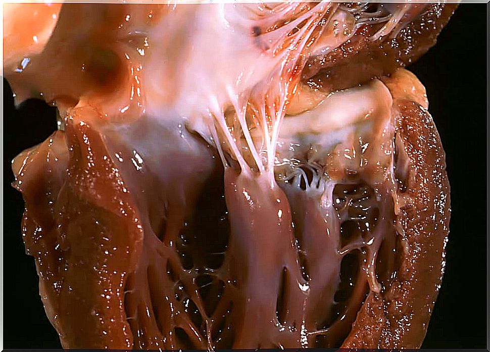
Also known as the heart or myocardium, the heart muscle is an involuntary contraction muscle located in the middle of the chest. This is one of the three types of muscle that exist in the human body together with the smooth (characteristic of the viscera) and the skeletal, which forms the locomotor system.
The cells that make up the heart muscle contain a single nucleus, which is responsible for pumping all the blood through the circulatory system through the process of contraction. In turn, this muscle is able to function without the need for nerve stimulation and it decides when it contracts and when it dilates.
As this process is of vital importance for the human body, it is convenient to know how it works and the different phases that have to happen for it to occur. Therefore, we explain it to you below in more detail.
What is the heart muscle made of?
The heart muscle is also capable of contracting, but what differentiates it from others is that it can respond to the information offered by the tissues thanks to the fact that it is composed of:
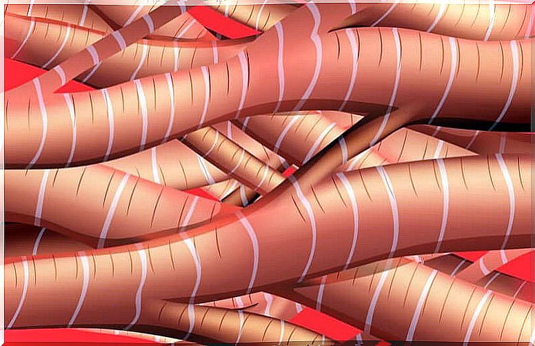
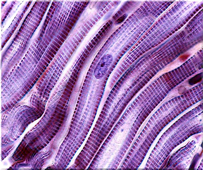
Striations
The heart muscle has striae and, according to the following article published by the University of Leeds, they are produced by segments of thick interlaced proteins (actin and myosin).
These proteins are also in skeletal muscle, but the difference between these two types is in the placement of the fibers. While in skeletal muscle they are in a linear and parallel position, in cardiac the fibers intertwine irregularly.
T tubules
T tubules are extensions and branches that are in the cell membrane that surrounds the muscle.
The main difference with respect to the skeletal muscle is and n heart, the T tubules are larger and wider. Likewise, the cells that make up its tissue also have fewer T tubules than those of the skeletal one.
Interlayer discs
They are the systems that unite the cardiac muscle cells. These structures are in the regions of the membrane where the ends of two cells meet.
How is the contraction of the heart muscle carried out?
The process between cardiac excitation and contraction is made up of a series of events based on the action potential. This process is important, as it allows the heart to beat in a controlled way and without the need for external nerve stimulation.
Coupling of the heart muscle is the sequential result of the contraction of the heart muscles. In fact, according to an article published by expert Kathleen H. McDonough, it is capable of contracting between 60 and 100 times per minute, even when the body is at rest.
However, this speed can be altered by the sympathetic and parasympathetic nerves, which can vary the rate of the beat.
Phases of the cardiac contraction process
According to the following scientific article published in 2019 by the Encyclopædia Britannica , the three main phases that take place during the process of cardiac contraction are:
Initiation
The cells located that constitute what is known as the sinoatrial (SA) node allow the muscle to contract spontaneously. This structure is considered as the “pacemaker” of the heart and the onset of cardiac contraction and the rate of the beat depend on it.
The generated action potentials are spread across the muscle cell membrane in the form of pulses. These impulses pass from one cell to another through structures known as “gap junctions”. However, their speed will vary depending on the part of the heart where they are.
First the upper chambers (atria) contract, and shortly thereafter the lower chambers (ventricles) contract. This slight delay allows blood to be stored within the ventricles before they fully contract. In fact, if it didn’t exist, the heart would explode.
“Calcium-induced release of calcium”
Different regions of the membrane penetrate inside the cells. Calcium gradients lead to them that depolarize the membrane and initiate the transmission of the action potential generated in the “pacemaker” of the heart.
The increase in calcium in the cell activates a receptor called ryanodine type 2 (RyR2). This receptor is located on the membrane of the sarcoplasmic reticulum (SR) and retains calcium within the T tubules.
In conclusion, the activation of the sarcoplasmic reticulum causes more calcium to be released within the cell. For this reason, the process is called “calcium-induced calcium release (ICRC)”.
Muscle contraction
The increase in calcium within the cell due to the release of calcium from the sarcoplasmic reticulum allows indirect activation of actin and myosin fibers. This process is what gives rise to muscle contraction.
But how does this happen? Calcium binds to a protein present in the actin fiber filaments called troponin. After this binding, the troponin undergoes a change to, in turn, clump to the region furthest from the adjacent myosin fiber. For this reason, the junction between the actin and myosin heads is known as a “cross bridge”.
Adenosine triphosphate (ATP) (a molecule produced in the mitochondria) is used as an energy source for the mobilization of the myosin head. On the other hand, actin slides through the myosin filament and causes it to shorten in the muscle; which leads to shrinkage.
Termination of heart muscle contraction
The contraction of the heart muscle ends when the intracellular concentration of calcium decreases. Thus, troponin returns to its original state, blocking actin binding areas and preventing the formation of “cross bridges”.
On the other hand, the decrease in calcium concentration occurs due to the opening of ion transporters. These are present in the membrane of the sarcoplasmic reticulum; which is the one that stores the calcium to release it with the arrival of the next contraction and, thanks to the presence of stem cells, this occurs successively.
To this day, scientists continue to investigate the regenerative versatility of stem cells found in this tissue to develop therapies, but more studies are still needed in this regard. Until then, we hope this information has been helpful to you and that you have learned more about the heart muscle.